The Songjiang Research Institute boasts a dedicated Electrophysiological Facility, designed to support both internal and external researchers in their electrophysiological experiments. This platform plays a pivotal role in advancing basic scientific research, spurring innovative technological development, and fostering personnel training.
Initially, the electrophysiology platform is equipped with a comprehensive brain slice electrophysiology system. This includes tools such as the patch clamp, electrical mapping instruments, and an isolated heart perfusion system. This setup allows researchers to conduct diverse electrophysiological studies, from monitoring isolated brain slice electrophysiology to brain tissue separation, cell electrophysiology, and ion channel-oriented experiments. Furthermore, the facility will also house two multi-channel electrophysiological recording systems, proficient in tracking ECG, EMG, Spike, and other electrical signals.
Integral to the platform's success are its specialized technical personnel. They ensure meticulous management and maintenance of the instruments while also offering essential training, evaluations, and related services.
1. Eeg electrophysiological system

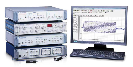
The Brain Patch Clamp Test stands as a benchmark functional experimental system, finding extensive application in both the neurological and cardiovascular research realms. The primary purposes of this system can be outlined as follows:
Signal Recording: The system captures the discharge (comprising current and voltage fluctuations) emanating from individual cells, tissue slices, or living organisms. Such recordings, particularly of current and voltage variations, facilitate the study of membrane ion channel protein functionality.
Stimulus Application: Depending on the researcher's objectives, this system can deliver specific stimuli to the sample—be it a single cell, tissue slice, or a whole organism. Recording the sample's response to various stimuli helps achieve the research goals.
Central to this system is a series of components:
Patch Clamp Amplifier: Among the system's components, the dual probe patch clamp amplifier is quintessential.
Data Converter: This device transitions the analog signals captured by the amplifier into digital formats, suitable for subsequent analysis and storage. Concurrently, it has the capability to transform artificial command signals from a digital back to an analog state, which can then be applied directly to the cell.
Data Acquisition and Analysis Software: This software ensures that the processed signals are recorded and securely stored on a computer for in-depth analysis. Additionally, it allows researchers to predetermine recording modes and devise specific stimulus commands. These are then dispatched to the data converter and amplifier for implementation.
In essence, the Brain Patch Clamp Test offers an intricate, yet robust, means to explore the intricacies of cellular functionality in response to various stimuli.
This system can record almost all electrophysiological signals, the main ones are:
(1) Action potential (Spike)
(2) Electromyography (EMG)
(3) Electroophthalmogram (EOG)
(4) Excitatory postsynaptic currents (EPSCs)
(5) Excitatory postsynaptic potentials (EPSPs)
(6) Inhibitory postsynaptic currents (IPSCs)
(7) Inhibitory postsynaptic potentials (IPSPs)
(8) Tiny excitatory potential (Minis)
(9) Long-term Enhancement (LTP)
(10) Long-term Inhibition (LTD)
(11) Fluorescent staining ratio
(12) Peak potential string
(13) Synaptic network
2. Multi-channel neural electrophysiological experimental system

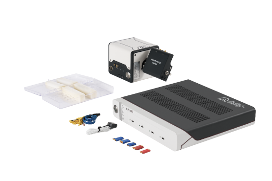
The alphaRS system is designed for advanced in vivo multi-channel electrophysiological data acquisition and stimulation. By implanting a single electrode or a microelectrode array, it captures real-time extracellular action potential signals (spikes) and local field potential signals (Local Field Potential) from neurons of either head-fixed or awake, freely moving animals. Once these signals undergo amplification and analog-to-digital conversion via the Headstage, the data is relayed to the alphaRS main unit for processing and then displayed.
Key Features of alphaRS:
High Channel Capacity: The system boasts up to 512 individual acquisition channels. Moreover, combining multiple alphaRS units allows for simultaneous data collection across a minimum of 2048 channels.
Integrated Stimulation Source: A built-in stimulus source eliminates the need for an external stimulator. With the same software interface as data collection, it offers a seamless one-click stimulus release.
Impedance Test Functionality: The built-in impedance test is integrated into the same software interface as the data acquisition, facilitating a one-click in vivo impedance test.
Minimal Delay: There's less than a 1.5ms delay in switching from stimulus to record on the same channel.
Spike Sorting Capabilities: The system supports online spike sorting, leveraging an 8-point matching SSQ algorithm. Each channel can be tailored to categorize up to four spike waveform templates.
Software Compatibility: The system is compatible with the NeuroExplorer data analysis software, accommodating both online and offline data analyses.
Robust Processing Power: Furnished with an eight-core DSP chip, the alphaRS system realizes high-speed data processing and rapid online data analysis directly within the main unit.
Built-in Acquisition and Stimulation Capabilities:
Distinctively, the alphaRS electrophysiological acquisition system is embedded with a stimulation function. Without relying on external stimulators, it can achieve full-channel acquisition and stimulation for up to 256 channels. The data acquisition interface permits one-click stimulation, and every channel is dual-purposed for both data acquisition and electrical stimulation.
In sum, the alphaRS system offers a comprehensive solution for researchers aiming to uncover intricate neuronal activity patterns and the underpinning neural mechanisms.
This device can record the following signal types:
(1) Extracellular high-frequency action potential of SPIKE neurons
(2) Local field potential in the brain region where the LFP electrode is located
(3) EMG electromyography recording
(4) ECoG cortical EEG recording
(5) EEG electrical signal recording
(6) Raw RAW wide-band signal


